 |
Darkfield |
|---|
 |
Darkfield |
|---|
Darkfield
Metallographers infrequently use darkfield (also called dark ground) illumination. Only a few phases are best examined with darkfield. In copper alloys, such as tough pitch copper, the copper oxide phase lights up vividly when viewed with darkfield, but the copper sulfide phase does not. With brightfield illumination, both phases look rather alike.
Darkfield, as with DIC, may reveal features not visible with brightfield illumination. Darkfield and DIC both generate improved image contrast for many microstructures. The self-luminous effect produced by darkfield offers increased resolution and visibility over brightfield.
Darkfield collects the light that is scattered by roughness, cracks, pits,
etch steps, or "ditches", etc. Hence, it is quite useful in the study of
grain structures. Figure 5 shows the grain structure of Waspaloy, a nickel-based
superalloy, in the solution annealed and double-aged heat treatment condition
after etching with glyceregia.
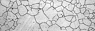
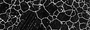
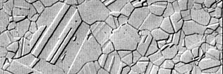
| Figure 5. Austenitic grain structure of a Waspaloy, a nickel-based superalloy, etched with glyceregia and viewed with (top) brightfield, with (middle) darkfield; and, with (bottom) differential interference contrast illumination. (IOOX) |

|

|
|

|
Figure 5 (top) shows the structure viewed with brightfield revealing a
partially developed grain structure and some faint annealing twins (the faint
parallel lines or bands within the grains). Figure 5 (middle) shows the same
area viewed with darkfield illumination. Basically, the contrast is reversed,
but all of the grain and twin boundaries are much more clearly revealed.
Also, we see some coarse precipitates within the grains (the fine, white
dots). Figure 5 (bottom) shows the same area examined with DIC which reveals
the grain structure well. This shows how the etchant has produced selective
dissolution to reveal the structure.
As a final example of the benefit of using alternate illumination modes, Figure 6 shows the microstructure of eutectoid aluminum bronze (Cu - 11.8 wt. % Al) heat treated to form martensite.
Go To: |
[Previous Page] |
[FAQ] |
[Home] |
[ Vendors] |
|---|